PSY3309
Behavioral Neuroscience


Dr. Smith
Florida Southern College
Fornix -Lateral View
Disclaimer: All structures that have been covered in the lab manual so far can be tagged on a test. This webpage covers the majority of the structures that can be tagged on this view, however, please note that there may be other structures not listed here that can be tagged on the fornix view.
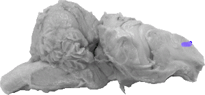
Anterior Horn of the Lateral Ventricle
The anterior horn of the lateral ventricle is the front boundary of the entire ventricle, and its a space that falls directly behind the genu of the corpus callosum.

Anterior Lobe
The anterior lobe of the cerebellum is the front central portion of the structure, and it is separated from the posterior lobe via the fissura prima.
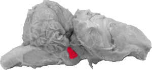
Brachium Pontis
The brachium pontis is the lateral extension of the pons, and, on this view projects upward into the cerebellum (flocculus).
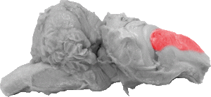
Caudate Nucleus
The caudate nucleus is a large area of grey matter that falls between the genu of the corpus callosum and the fimbria. It makes up a major portion of the basal ganglia system.
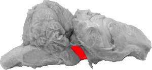
Cerebral Peduncle
The cerebral peduncle is the ventral portion of the mid brain and is located directly in front of the pons and brachium pontis.

Corpus Cerebelli
The corpus cerebelli represents the majority of the lateral cerebellum.
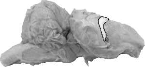
Fimbria
The fimbria is a web-like band of fibers rosteral to the hippocampus as it wraps around the thalamus (not seen in this view).
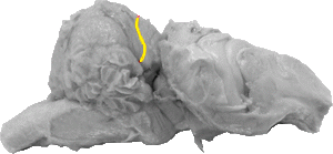
Fissura Prima
The fissura prima is the line that separates the anterior and posterior lobes of the cerebellum.
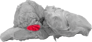
Flocculus
The flocculus is the ventro-lateral lobule of the cerebellum that falls directly above the trapezoid body, the trigeminal nerve, and the brachium pontis.

Fornix
The fornix can barely be seen on this view, but it would be labeled as a white central strip that is directly rosteral to the splenium.The fimbria and the fornix fibers are projections that arise from this structure.

Fornix Fibers
The fornix fibers cover the banana shaped hippocampus, which runs around the thalamus (not pictured). These fibers wrap around the hippocampus like the peel of a banana.
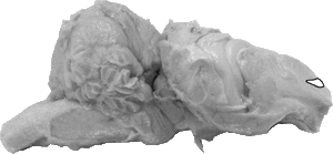
Genu of the Corpus Callosum
The genu of the corpus callosum marks the front of the entire fiber bundle, and it serves as a rosteral boundary for the anterior horns of the lateral ventricle as well as the caudate nucleus.

Hippocampus
The hippocampus is the grey matter that wound comprise the banana like structure going down the side of the brain (around the thalamus which is not pictured).
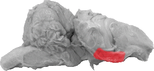
Hippocampal Gyrus
The hippocampal gyrus marks the ventral boundary of the forebrain, and it is separated from the hippocampus by the inferior horn of the lateral ventricle.
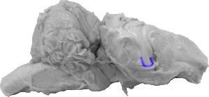
Inferior Horn of the Lateral Ventricle
The inferior horn is the bottom most boundary of the lateral ventricle and it can be seen as a separation between the hippocampus/fimbria and the hippocampal gyrus.
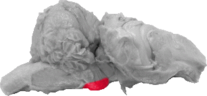
Pons
The pons is a large ventral protrusion between the trapezoid body and the cerebral peduncle.

Posterior Horn of the Lateral Ventricle
The posterior horn of the lateral ventricle is the space behind the hippocampus and is the very back boundary of the entire lateral ventricle.
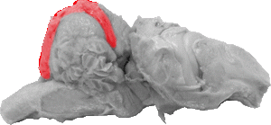
Posterior Lobe
The posterior lobe represents the majority of the central cerebellum on this view and it is separated from the anterior lobe of the cerebellum by the fissura prima.

Pyramid
The pyramid is the ventro-medial fiber tract of the medulla.
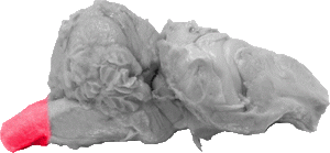
Spinal Cord
The spinal cord represents the very caudal portion of this view.
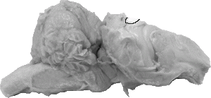
Splenium of the Corpus Callosum
The splenium of the corpus callosum can be seen as white matter that is directly behind the very top of the hippocampus.

Superior Colliculus
The superior colliculus is a protrusion of matter on the very top of the cerebral peduncle. It represents the dorsal portion of the midbrain.
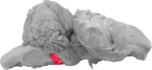
Trapezoid Body
The trapezoid body is a band of matter that is ventral to the flocculus and caudal to the trigeminal nerve. It is the very front of the medulla.

Trigeminal Nerve
The trigeminal nerve is a large projection of nerve fibers between the trapezoid body and the brachium pontis.