PSY3309
Behavioral Neuroscience


Dr. Smith
Florida Southern College
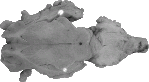
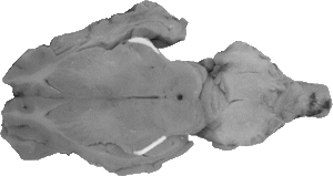
Horizontal -Lower View
Disclaimer: All structures that have been covered in the lab manual so far can be tagged on a test. This webpage covers the majority of the structures that can be tagged on this view, however, please note that there may be other structures not listed here that can be tagged on the horizontal view.
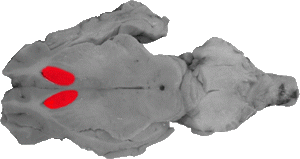
Anterior Horn of the Lateral Ventricle
The anterior horn of the lateral ventricle is the space directly in front of the caudate nucleus (is should be noted that on most lower views the genu of the corpus callosum might not be seen).
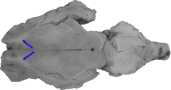
Anterior Limb of the Internal Capsule
The anterior limb of the internal capsule can be faintly seen as white projections lateral to the caudate nucleus.

Anterior Lobe of the Cerebellum
The anterior lobe of the cerebellum can be seen directly caudal to the tectum and the inferior colliculi. It is usually the only visual lobe of the cerebellum in a lower horizontal cut.

Arbor Vitae
The arbor vitae represent the white matter throughout the middle of the cerebellum.
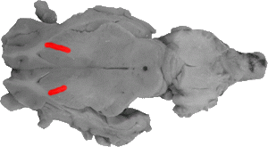
Caudate Nucleus
The caudate nucleus is the grey matter that runs laterally and caudally to the anterior horns of the lateral ventricle. On this view it is the only discernible part of the basal ganglia system.
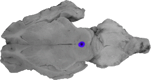
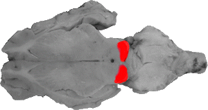
Cerebral Aqueduct
The cerebral aqueduct is the small circular opening in the center of the midbrain and can be seen in most lower horizontal cuts.
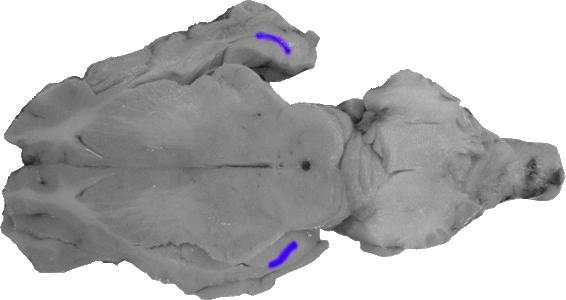
Fimbria
Similar to the upper horizontal section, the fimbria is a small white tip on each hippocampus.
Hippocampus
Similar to the upper horizontal cut the hippocampus is an area of grey matter that is caudal and lateral to the back of the thalamus.

Hippocampal Gyrus
On a lower horizontal cut, the grey matter lateral and caudal to the hippocampus is the hippocampal gyrus.
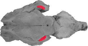
Inferior Colliculus
On a lower cut the prominent feature should be the inferior colliculi, which are two small rounded masses that fall to each side of the anterior lobe of the cerebellum. It should be noted that the inferior colliculi are not touching and are separated by the back portion of the tectum.
Inferior Horn of the Lateral Ventricle
On a lower horizontal cut (denoted by a view of the inferior colliculi), the space behind the hippocampus is the inferior horn of the lateral ventricle.
Optic Tract
The optic tract goes into the lateral portions of the thalamus on this lower view. These fiber projections go directly into a part of the thalamus called the lateral geniculate nucleus.

Spinal Cord
The spinal cord marks the caudal portion of this view.
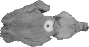
Tectum
On a lower horizontal cut the superior colliculus cannot be seen, and thus the white area covering the midbrain is in fact the tectum. It usually surrounds a small circular opening, known as the cerebral aqueduct.
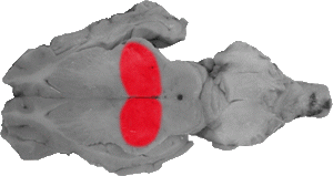
Thalamus
The thalamus represents large rounded masses in the middle of the view.

Third Ventricle
The third ventricle can be seen as a central space towards the back of the thalamus in this view.