PSY3309
Behavioral Neuroscience


Dr. Smith
Florida Southern College
Ventral View
Disclaimer: All structures that have been covered in the lab manual so far can be tagged on a test. This webpage covers the majority of the structures that can be tagged on this view, however, please note that there may be other structures not listed here that can be tagged on the ventral view.
Abducens Nerve
The abducens nerve (cranial nerve 6) is connected to the trapezoid body.

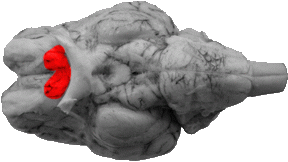
Anterior Perforated Substance
The anterior perforated substance is bound caudally by the olfactory bulb, medial to the medial olfactory gyrus/longitudinal fissure, laterally by the lateral olfactory stria and rostrally by the optic tract. These boundaries form a rhomboid shaped area.

Brachium Pontis
The brachium pontis (also called the middle cerebellar peduncle), is the lateral extension of the pons into the cerebellum. It can be located directly rostral to the trigeminal nerve (which is also located to the side of the pons).
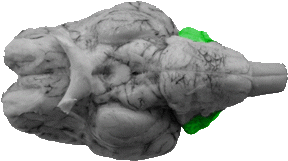
Cerebellum
On the ventral view much of the cerebellum is hidden. The only areas that can be seen on this view are the corpus cerebelli and the flocculus.
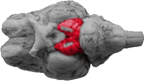
Cerebral Peduncle
The cerebral peduncle is a large bundle of axons that ipselaterally project between the cerebral cortex and the brainstem. These peduncles are rostral to the pons and its lateral extremities. It can also be noted that the peduncles are separated by the interpeduncular fossa and hosts the origination of the ocularmotor nerve.
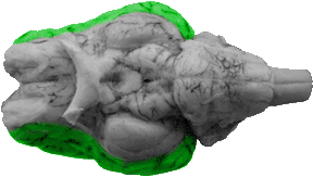
Cerebral Cortex
On the ventral view much of the cerebral cortex cannot be seen. However, parts of the frontal lobe, temporal lobe, occipital lobe, and insula can be distinguished.
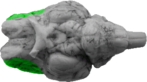
Frontal Lobe
Much of the frontal lobe cannot be seen on the ventral view. However, the inferior frontal gyrus can be found lateral (and dorsal) to the rhinal fissure at the very front of the brain.
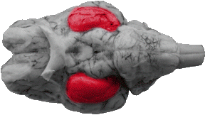
Hippocampal Gyrus
The hippocampal gyrus is the caudal portion of the pyriform lobe.

Infundibulum
The infundibulum (also called the pituitary stalk) is a projection or hole that is located directly caudal to the optic chiasm on the ventral view and is also directly rostral to the mammillary bodies. It is the site for which hypothalamic signals travel directly to the pituitary gland (for hormone secretion).

Insula
The insula is a small area of matter that is surrounded by the anterior and posterior rami of the lateral fissure, and the rhinal fissure. This area is now considered to be a major lobe of cortex whose function involves the mediation of taste.

Interpeduncular Fossa
The interpeduncular fossa is the medially deep grove that runs between the mammillary bodies and the pons. It is designed to separate the two cerebral peduncles.
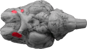
Lateral Olfactory Gyrus
The lateral olfactory gyrus is the rostral portion of the pyriform cortex.
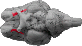
Lateral Olfactory Stria
The lateral olfactory stria is a white stripe that originates from the olfactory bulb and runs medially to the lateral olfactory gyrus.

Longitudinal Fissure
The longitudinal fissure can be found on the front one third of the ventral view. It is the line that separates the two medial olfactory gyri.

Mammillary Bodies
The mammillary bodies are projections that are surrounded by the infundibulum (rostrally) and the cerebral peduncles (laterally) and the interpeduncular fossa.

Medial Olfactory Gyrus
The medial olfactory gyrus is located right off the longitudinal fissure and is medial to the olfactory bulb and anterior perforated substance.

Occipital Lobe
The most caudal portion of the occipital lobe can be seen on the ventral view in which it's separated from the hippocampal gyrus by means of the caudal part of the rhinal fissure.
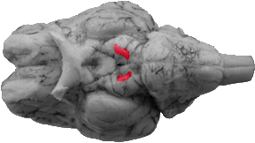
Oculomotor Nerve
The oculomotor nerve originates from the middle of the cerebral peduncle.
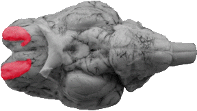
Olfactory Bulb
The olfactory bulb is an oval shaped structure located towards the rostral part of the brain. It is lateral to the medial olfactory gyrus and is medial to the rhinal fissure. The olfactory bulb directly receives sensory input from the nasal cavity (via the olfactory nerves), and it is critical for the processing of smell. It should be noted that a human olfactory bulb is considerably smaller than what is seen in the sheep.

Olive
The olive is a rounded elevation on the medulla that is caudal to the trapezoid body and lateral to the pyramid.

Optic Chiasm
The optic chiasm is the medial junction of the optic nerves (coming from the eyes) and the optic tracts ascending into the brain. It can be seen caudal to the back of the longitudinal fissure and rostral to the infundibulum. It is the site where visual information from each eye crosses to the opposite side of the brain.
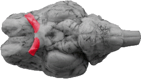
Optic Nerve
The optic nerve (cranial nerve number 2) is a projection from the eyes to the optic chiasm.
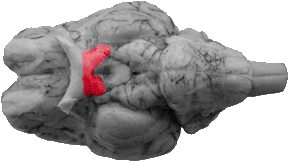
Optic Tract
The optic tract is a fiber bundle that runs caudally and laterally from the optic chiasm into the brain (under the hippocampal gyrus).

Pons
The pons is a large rectangular elevation that is caudal to the cerebral peduncles and the interpeduncular fossa. It has on each side a lateral projection of the pons (known as the brachium pontis, or middle cerebellar peduncle) that projects into the cerebellum.

Pyramid
The pyramid is a long rectangular elevation just lateral to the mid-line of the medulla (also called the ventral median fissure). It starts at the pons and runs caudally to the spinal cord. These elevations are caused by the pyramidal, or corticospinal tracts (an important fiber system for voluntary movement), and they form a 'T' with the trapezoid body.
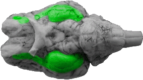
Pyriform Lobe
The pyriform lobe is part of the rhinencephalom, and it consists of the lateral olfactory gyrus and the hippocampal gyrus. This area is involved with smell perception.

Rhinal Fissure
The rhinal fissure is a line that separates the cerebral cortex from the arche cortex (the primitive cortex in mammals). It runs the entire length of the cerebral cortex and is lateral to the olfactory bulb, the lateral olfactory gyrus, and the hippocampal gyrus.

Spinal Accessory Nerve
The spinal accessory nerve runs laterally to the medulla and spinal cord.
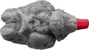
Spinal Cord
The spinal cord begins right after the caudal medulla. On the ventral surface one can discern the continuation of the pyramidal tracts.
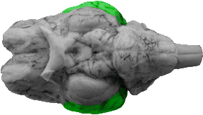
Temporal Lobe
In higher level mammals, the temporal lobe is separated from the frontal lobe by the lateral fissure. In the sheep, branches of the lateral fissure separate the insula from the temporal lobe as well.
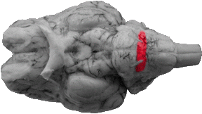
Trapezoid Body
The trapezoid body is a rectangular area that is directly caudal to the pons. The abducens nerve can be seen to project off this structure, which also forms a 'T' with the trapezoid body.
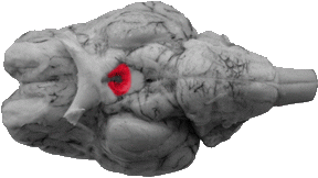
Tuber Cinereum
The tuber cinereum is the lateral area around the infundibullum. Technically, it is the very bottom surface of the hypothalamus.
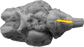
Ventral Median Fissure
The ventral median fissure is a line that separates the two pyramids along the middle of the medulla.